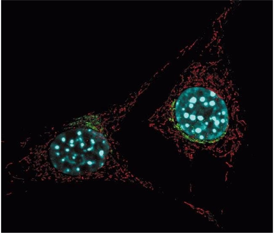
MitoTracer™ Red FM (Known as MitoTracker® Red FM, Trademark of Thermo Fisher Scientific) is an red-fluorescent dye that stains mitochondria in live cells and its accumulation is dependent upon membrane potential. The dye is not well-retained after aldehyde fixation.
Reference:
Frequently asked questions (FAQs)
It looks like my Mitotracker™ dye is staining more than just the mitochondria. Why?
Specifications:
Excitation/Emission:
580/644 nm
Shipping Condition:
Ambient
Storage Conditions:
-20ºC, protect from light
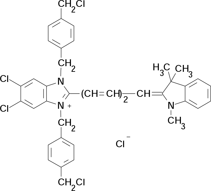
Molecular Formula:
C39H36Cl5N3
Molecular Weight:
724
CAS Number:
–
Protocol (PDF):
C054
MSDS (PDF):
MSDS-C054
COA (PDF):
C054
J Biol Chem (2006) 281:10374-10380
Genes Dev (2012) 26:2027-2037
Product usage: Proliferation, DNA Synthesis, Click-iT EdU, Imaging, in vivo
J Biol Chem (2012) 287:23615-23625
Product usage: Lysotracker, ER-Tracker, Mitotracker, Autophagy, Imaging
This is typically a result of using too high of a concentration of the MitoViewr™ dye. Most organic dyes are used in the low micromolar range. The MitoView™ dyes are used at a much lower concentration, around 50–200 nanomolar. Higher concentrations can cause background fluorescence and non-mitochondrial staining.
Home » Cell Structure Probes » MitoTracer™ Red FM
MitoTracer™ Red FM
Introduction
To Order
Documents
Iuso A, Scacco S, Piccoli C, Bellomo F, Petruzzella V, Trentadue R, Minuto M, Ripoli M, Capitanio N, Zeviani M, Papa S
Nagaraj R, Gururaja-Rao S, Jones KT, Slattery M, Negre N, Braas D, Christofk H, White KP, Mann R, Banerjee U,
Zhao T, Huang X, Han L, Wang X, Cheng H, Zhao Y, Chen Q, Chen J, Cheng H, Xiao R, Zheng M,
Related Products
Contact Us
ABP Biosciences
405 E Gude Dr, STE 214
Rockville, MD 20850
Service Hotline
Tel: 301-658-7993
E-mail: info@abpbio.com
Copyright@ 2026 ABP Biosciences, LLC. All Rights Reserved.
