Our Cell-Quant™ No Wash Cell Proliferation Assay Kit provides an accurate microplate–based fluorescence method for counting cells in a population, based on cellular DNA content. Our Cell-Quant™ Assay provides a very convenient workflow for high-throughput screening of cell proliferation and cytotoxicity–no washes, cell lysis, temperature equilibrations, or radioactivity required. The Cell-Quant™ Assay has a linear detection range from less than 100 to 20,000 cells per well in most cell types. The add-mix-read assay format makes it ideal for HTS applications. The assay can be completed in one hour, and the signal is stable for several hours, affording additional work flow convenience.
Features
Specifications
Applications
Cell proliferation and cytotoxicity analysis
Reference:
Abraham VC, Towne DL, Waring JF, Warrior U, Burns DJ,
Activator of G protein signaling 3 promotes epithelial cell proliferation in PKD.
Nadella R, Blumer JB, Jia G, Kwon M, Akbulut T, Qian F, Sedlic F, Wakatsuki T, Sweeney WE, Wilson PD, Lanier SM, Park F,
Tan F, Naciri M, Al-Rubeai M,
Platform:
Fluorescence plate reader
Detection Method:
Fluorescent
Ex/Em:
490/520 nm
Product Size:
1000 assays
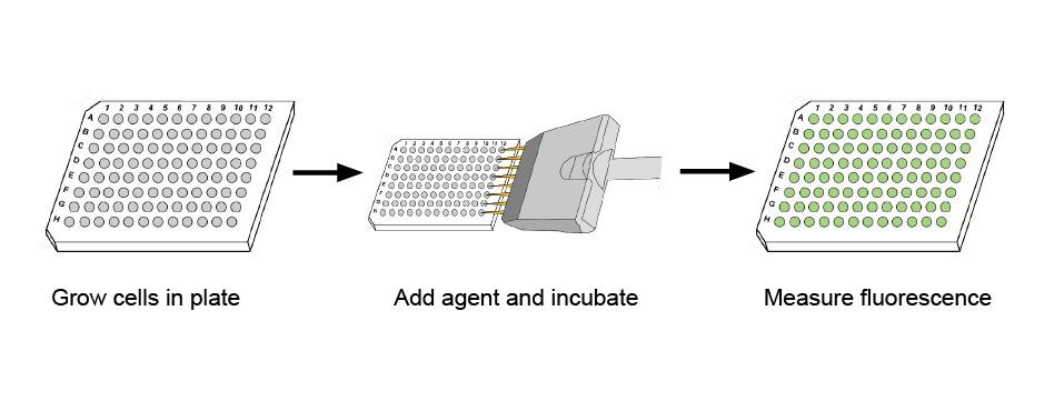
Figure 1. Cell-Quant™ No Wash Cell Proliferation Assay Workflow
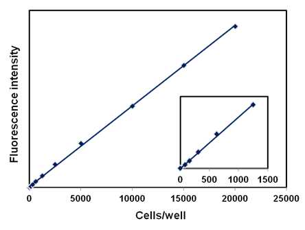
Figure 2. Quantitation of CHO cells using the Cell-Quant™ No Wash Cell Proliferation Assay Kit. Fluorescence measurements were made using a microplate reader with excitation at 485 nm and emission detection at 530 nm. The linear range of the assay under these conditions is from 100 to 20,000 cells per 100 µl sample. The inset shows the linearity that can be obtained at very low numbers of cells.
Protocol (PDF):
A011
MSDS (PDF):
MSDS-A011
J Biomol Screen (2008) 13:527-537
J Am Soc Nephrol (2010) 21:1275-1280
Biotechnol Bioeng (2011) 108:454-464
Home » Cell Proliferation and Viability » Cell-Quant™ No Wash Cell Proliferation Assay Kit
Cell-Quant™ No Wash Cell Proliferation Assay Kit
Introduction
To Order
Documents
Related Products
Contact Us
ABP Biosciences
405 E Gude Dr, STE 214
Rockville, MD 20850
Service Hotline
Tel: 301-658-7993
E-mail: info@abpbio.com
Copyright@ 2026 ABP Biosciences, LLC. All Rights Reserved.
