JC-1 Mitochondrial Membrane Potential Detection Kit provides a quick and reliable method to detect the mitochondrial membrane potential changes in apoptotic cells using flow cytometry. The loss of mitochondrial membrane potential is a hallmark for apoptosis. The JC-1 Assay Kit uses a unique cationic dye (5,5’,6,6’- tetrachloro-1,1’,3,3’-tetraethylbenzimidazolylcarbocyanine iodide) to signal the loss of mitochondrial membrane potential. JC-1 exhibits potential-dependent accumulation in mitochondria, indicated by a fluorescence emission shift from green (529 nm) to red (590 nm). Consequently, mitochondrial depolarization is indicated by a decrease in the red/green fluorescence intensity ratio. In healthy cells, the JC-1 accumulates in the mitochondrial to form J-aggregates, which become red fluorescence. In apoptotic cells, the JC-1 cannot accumulate within the mitochondria and remains in the cytoplasm in monomeric form to show green fluorescence.
Features
Reference
Specifications
Platform:
Fluorescence Microscopy, Flow Cytometry, Plate Reader
Detection Method:
Fluorescent
Ex/Em:
514/529, 590 nm
Product Size:
100 assay
Storage Conditions:
-20 ℃, protect from light
Shipping Condition:
Room Temperature
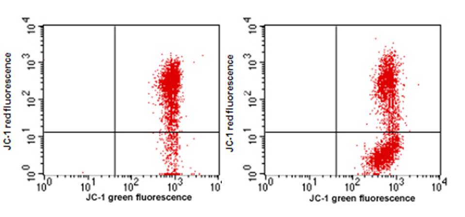
Figure 1. Detection of cell apoptosis with JC-1 Mitochondrial Membrane Potential Detection Kit. Jurkat cells treated with 10 μM camptothecin for 4 hr (right panel) or untreated (left panel). Cells were stained with JC-1 reagent, then analyzed by flow cytometry.
Protocol (PDF):
A048
MSDS (PDF):
Component A
Component B
COA (PDF):
COA-A048
Mol Cancer Ther (2011) 10:1276-1288
J Biol Chem (2004) 279:54783-54792
J Biomol Screen (2008) 13:185-193
Related Products
Home » Cell Apoptosis Assay » JC-1 Mitochondrial Membrane Potential Detection Kit
JC-1 Mitochondrial Membrane Potential Detection Kit
Introduction
To order
Documents
Zhou H, Marks JW, Hittelman WN, Yagita H, Cheung LH, Rosenblum MG, Winkles JA,
Rodolfo C, Mormone E, Matarrese P, Ciccosanti F, Farrace MG, Garofano E, Piredda L, Fimia GM, Malorni W, Piacentini M
Winter SS, Lovato DM, Khawaja HM, Edwards BS, Steele ID, Young SM, Oprea TI, Sklar LA, Larson RS,
Related products
Contact Us
ABP Biosciences
405 E Gude Dr, STE 214
Rockville, MD 20850
Service Hotline
Tel: 301-658-7993
E-mail: info@abpbio.com
Copyright@ 2026 ABP Biosciences, LLC. All Rights Reserved.
