Cell Structure Probe
To view this email as a webpage, click here
|

|
|
Dear Scientist,
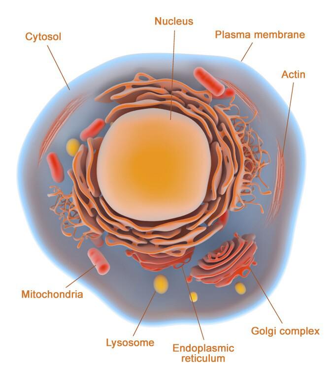
ABP Biosciences is proud to introduce a diverse selection of cell structure probes to specifically stain from organelle and membrane to whole cell. Our cell structure probes are widely used as counterstains to identify the location of specific proteins and targets of interest within the cell.
Special promotion: 30% off. Offer is valid until 11/31/2023.
Read on to find out more about cell structure probes.
|
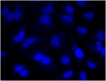
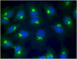
Nuclei Stains
|
ABP Biosciences offers a selection of nucleic acid stains to stain live or dead cells/tissues, thereby providing a means to locate the nucleus and follow nuclear changes throughout cellular processes, from mitosis to apoptosis.
See the Nucleic Acid Stains Selection Guide >
|
|
|
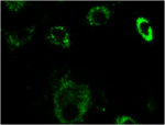
Membrane Stains
|
The cell membrane separates the cell from the extracellular environment and plays important roles in cell signalling pathways, as well as in ionic homeostasis. The membrane stains are useful markers for highlighting cell boundaries. ABP Biosciences offers a selection of lipophilic dyes that may be used as plasma membrane stains
See the Membrane Stains Selection Guide >
|

|
|
Cytosol Stains
|
Cytosol stains are useful probes to monitor cell morphology and cytosolic localisation in cell proliferation and viability studies. ABP Biosciences offers a selection of cell tracer dyes which can freely diffuse through the membranes of live cells, and become membrane-impermeant after loading.
See the Cytosol Stains Selection Guide >
|
|
|
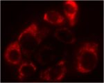
Mitochondrial Stains
|
ABP Biosciences has developed a series of mito-tracker probes which can be selectively sequestered by mitochondria in live cells based on the mitochondrial membrane potential for loading. ABP Biosciences' mito-tracker probes have a wide range of emission spectra for multicolour imaging.
See the Mitochondrial Stains Selection Guide >
|
|
|
Lysosome Stains
|
Lysosomes are membrane-bound cell organelles found in most animal cells. ABP Biosciences offers LysoView probes that appear to accumulate in acidic organelles. The LysoView probes exhibit a pH-dependent increase in fluorescence intensity upon acidification.
See the Lysosome Stains Selection Guide >
|
|
|
Golgi Stain
|
The Golgi contains a set of glycosylation enzymes that attach various sugar monomers to proteins as the proteins move through the apparatus. ABP Biosciences offers a cell-permeant probe that selectively stains the Golgi complex for lipid metabolism and trafficking studies.
See the Golgi Stain Selection Guide >
|
|
|
Endoplasmic Reticulum Stain
|
The endoplasmic reticulum (ER) has a central role in lipid and protein synthesis, protein chaperoning and folding, and calcium homeostasis. ABP Biosciences offers a cell-permeant probe that selectively stains the ER in live cells based on the probe concentration.
See the Endoplasmic Reticulum Selection Guide >
|

|
|
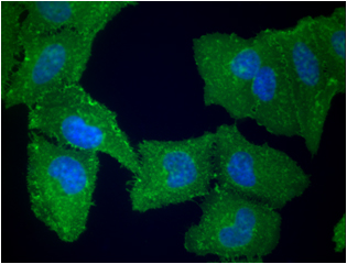
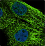
Cytoskeleton Stains
|
The cytoskeleton plays important roles in organelle transport, cell division, motility, and signaling, thereby making it central to both cell health and disease processes. ABP Biosciences offers a selection of phalloidin conjugates to label actin in fixed and permeabilised cells.
See the Cytoskeleton Stains Selection Guide >
|
|
Related Products
|
|
|
|

|
Contact us:
Phone: 1-301-658-7993
Email: info@abpbio.com
Website: www.abpbio.com
|
7040 Virginia Manor Road, Beltsville, MD 20705, USA
|
|
Click Here to Unsubscribe